Advanced Aortic Imaging for Clinical and Translational Research (AAI-CTR) Group
Pioneering precision imaging for cardiovascular health
The AAI-CTR Group, part of the Cardiovascular Imaging Section in the Department of Radiology at the University of Wisconsin–Madison School of Medicine and Public Health, develops and applies advanced CT and MRI–based tools to better detect, measure, and manage aortic disease.
By integrating quantitative imaging with clinical data, our research aims to improve diagnosis, risk prediction, and surgical planning for patients with aortic and aortic valve disorders. Our multidisciplinary team of radiologists, engineers, computer scientists, and surgeons collaborates to create reproducible imaging pipelines, validate new biomarkers, and translate innovations into clinical care.
Current projects include NIH-funded studies using 4D Flow MRI to identify hemodynamic factors associated with complications in aortic dissection, as well as the development of vascular deformation mapping (VDM)—a 3D technique for quantifying aortic growth created in partnership with industry collaborators. Through this work, we aim to advance understanding of aortic disease and enhance outcomes for patients.
Recent News & Events
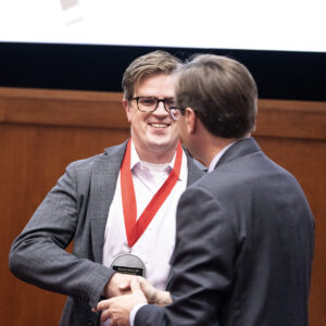
Dr. Nicholas Burris named inaugural David A. Bluemke, MD, PhD Professor of Radiology
The AAI-CTR Group celebrates Dr. Burris’s inaugural appointment recognizing leadership in advanced aortic imaging and translational radiology at UW–Madison.
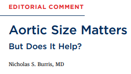
“Aortic Size Matters: But Does It Help?”
Published August 2025, Dr. Burris’s editorial comments on clinical interpretation of aortic dimensions and discusses the study “Ascending Aortic Dimensions and Body Size.”
First full-lab, in-person meeting at UW–Madison
Since Dr. Burris joined UW–Madison, the AAI-CTR Group reached a key milestone this October 2025—our first full-team, in-person meeting to kick off collaborative projects in advanced aortic imaging.
Dr. Burris’ publications on PubMed, Scholar Page
Select Publications
- Wagner CM, Marway PS, Ferrel MN, et al Sex differences in ascending aortic diameter at the time of acute type A aortic dissection Heart Published Online First: 12 October 2025. https://doi.org/10.1136/heartjnl-2025-325984
- Braet DJ, Baker TJ, Eliason JL, Figueroa CA, Burris NS. Assessing differences in growth and shape between symptomatic and asymptomatic abdominal aortic aneurysms. Quant Imaging Med Surg. 2025;15(7):5955–5968. https://doi.org/10.21037/qims-2024-2985
- Marway PS, Campello Jorge CA, Ahmad RA, Tjahjadi N, Patel HJ, Yang B, Burris NS. Distal anastomotic new entry tears predict long-term outcomes after hemi-arch repair for DeBakey I aortic dissection. Eur J Cardiothorac Surg. 2025;67(6):ezaf170. https://doi.org/10.1093/ejcts/ezaf170
- Tjahjadi NS, Campello Jorge CA, Marway PS, Knauer HA, Hazenberg C, van Herwaarden J, Figueroa CA, Patel HJ, Burris NS. Assessment of aortic arch involvement in ascending thoracic aortic aneurysm by three-dimensional growth mapping using CT angiography. Eur Radiol. 2025;35(6):3508–3518. https://doi.org/10.1007/s00330-024-11239-9
- Tjahjadi N, Campello Jorge C, Marway PS, Kim T, Baker T, Hazenberg C, van Herwaarden JA, Figueroa CA, Patel HJ, Burris NS. Three-dimensional characterization of ascending aortic strain, motion and growth in patients undergoing thoracic endovascular aortic repair. JVS Vasc Sci. 2025;6:100293. https://doi.org/10.1016/j.jvssci.2025.100293
- Henry M, Campello Jorge CA, van Bakel PAJ, Knauer HA, MacEachern M, van Herwaarden JA, Teixidó-Tura G, Evangelista A, Jeremy RW, Figueroa CA, Patel HJ, Hofmann Bowman M, Eagle K, Burris NS. Thoracic aortic aneurysm growth rates and predicting factors: a systematic review and meta-analysis. J Am Heart Assoc. 2025;14(7):e038821. https://doi.org/10.1161/JAHA.124.038821
- Marway PS, Campello Jorge CA, Tjahjadi N, Baker TJ, Mistelbauer G, Baeumler K, Hinostroza V, Higashigaito K, Mastrodicasa D, Masotti M, Nordsletten D, Patel HJ, Fleischmann D, Burris NS. Early three-dimensional growth in uncomplicated type B aortic dissection is associated with long-term outcomes. J Vasc Surg. 2025;81(1):75–84.e2. https://doi.org/10.1016/j.jvs.2024.08.059
- Braet DJ, Baker TJ, Delbono L, Spahlinger G, Graham N, Arora A, Figueroa CA, Eliason JL, Burris NS. Three-dimensional characterization of sex differences in abdominal aortic aneurysm progression via vascular deformation mapping. Sci Rep. 2024;14(1):24215. https://doi.org/10.1038/s41598-024-75334-z
- Katakol S, Baker TJ, Bian Z, Lu Y, Spahlinger G, Hatt CR, Burris NS. Fully automated pipeline for measurement of the thoracic aorta using joint segmentation and localization neural network. J Med Imaging (Bellingham). 2023;10(5):051810. https://doi.org/10.1117/1.JMI.10.5.051810
- Burris NS, Bian Z, Dominic J, Zhong J, Houben IB, van Bakel TMJ, Patel HJ, Ross BD, Christensen GE, Hatt CR. Vascular deformation mapping for CT surveillance of thoracic aortic aneurysm growth. Radiology. 2022;302(1):218–225. https://doi.org/10.1148/radiol.2021210658
- Burris NS, Hoff BA, Kazerooni EA, Ross BD. Vascular deformation mapping (VDM) of thoracic aortic enlargement in aneurysmal disease and dissection. Tomography. 2017;3(3):163–173. https://doi.org/10.18383/j.tom.2017.00015
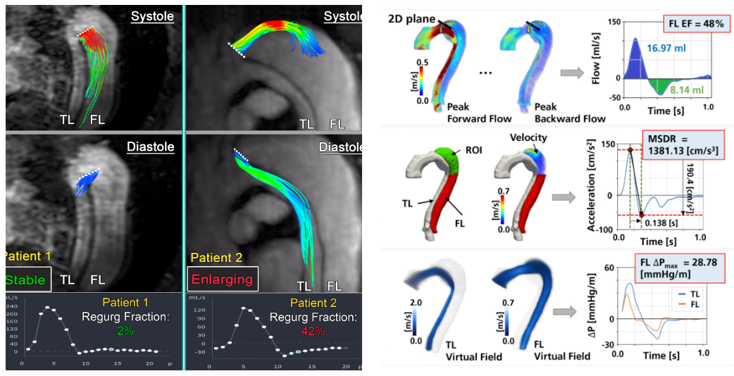
4D Flow in Aortic Dissection
4D Flow MRI is and advanced technique that allows for dynamic 3D quantification of blood flow in the aorta and other large vessels and has been applied broadly to study blood flow abnormalities through the body. Our group has specific interest in how the 4D Flow technique can be used to understand blood flow abnormalities in aortic dissection (AD), a disease that leads to two distinct channels for blood flow (true and false lumens), often displays complex anatomy and is associated with poor long-term outcomes in the chronic phase. Blood flow abnormalities in AD are not well understood, but our group has performed preliminary studies showing that there are unique aortic blood flow and pressure abnormalities in the false lumen of patients AD that can be used to understand which patients are at highest risk for aortic growth and need for surgical repair. This work has been supported by prior and current grants from the Radiologic Society of North America and the NIH National Heart Lung & Blood Institute (R01HL170059).
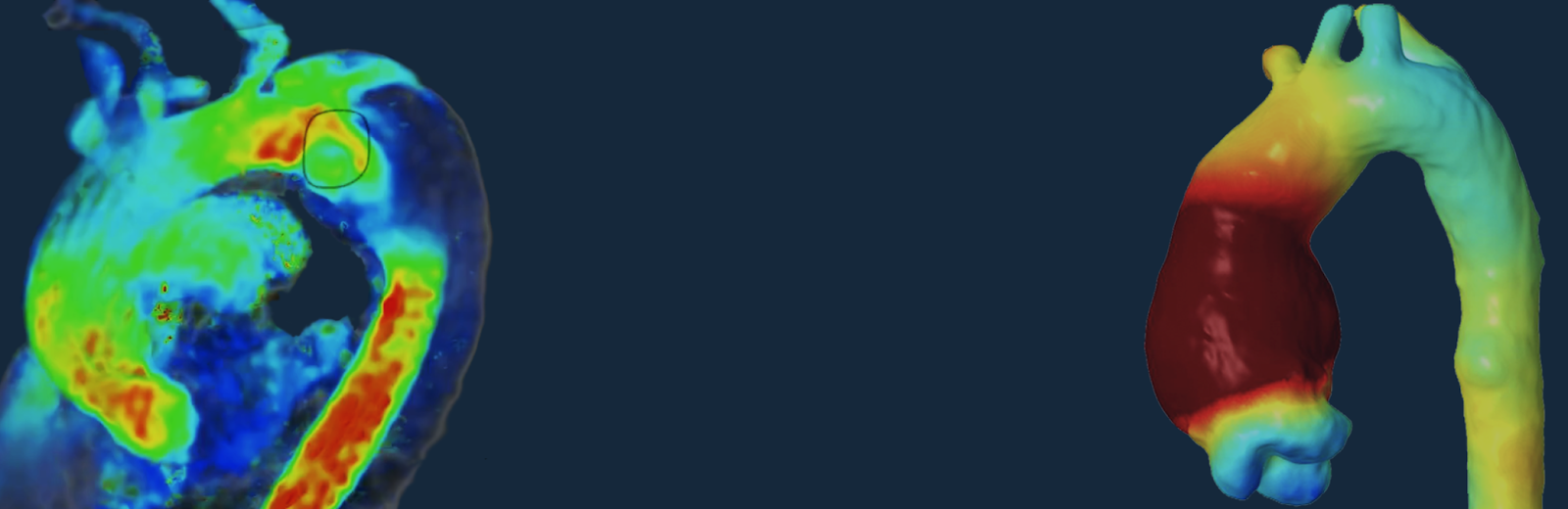
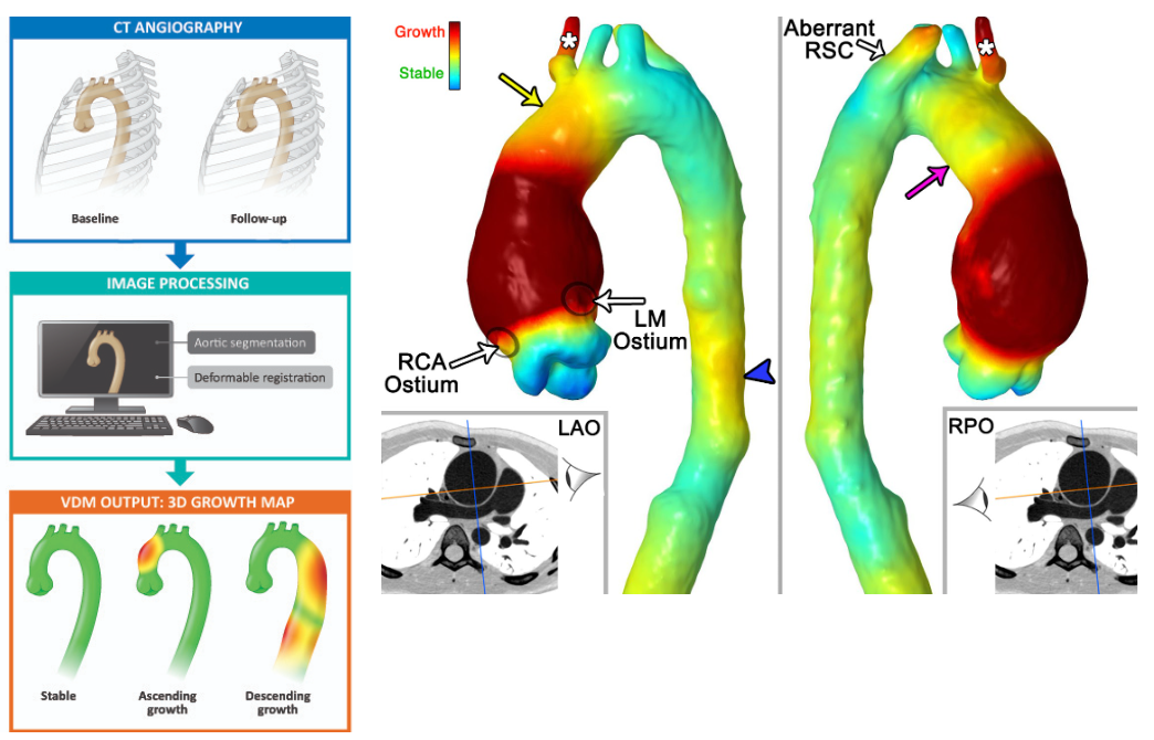
Vascular Deformation Mapping (VDM)
Vascular Deformation Mapping is an image analysis technique recently developed in our lab that takes routine gated computed tomography angiography (CTA) data and performs an analysis of 3-dimensional aortic growth in a matter that is more comprehensive and accurate than can be achieved with standard manual diameter-based measurements. We have validated this technique and are currently investigating VDM’s uses for better understanding growth patterns, predicting future growth & adverse events and assisting with surgical planning. VDM has been developed in conjunction with industry partners though the support of an NIH Small Business Innovation Research grant (R44HL145953)

Deep Learning for Automated Aortic Segmentation and Analysis
Segmentation and manual measurement of the aorta are time consuming tasks, even for expert readers, but are central components in most aortic analyses. These tasks can be greatly accelerated through image analysis techniques such as deep learning (DL). We are focused on developing and optimizing DL techniques to assist with aortic image analysis though a variety of tasks including automated aortic measurements and registration. The objective of this work is to make aortic image analysis more accurate, efficient and reproducible, and to advance the automation of other algorithms being developed in the lab.
Principal Investigator

Nicholas S. Burris, M.D.
Dr. Burris is the Principal Investigator of our Group. Having trained and held leadership roles at top institutions such as UCSF and the U-Michigan, he now leads our efforts at UW–Madison. As PI, he supervises all projects, ensuring that the work produced by the group meets the highest academic and scientific standards. The group is designed to balance close mentorship and oversight with academic freedom and innovation, so that every researcher can grow, contribute, and take ownership of their work.
Research Staff & Postdoctoral Associates

Gregory W. Spahlinger, Ph.D.
Greg is a research analyst and Python developer supporting AAI-CTR’s analytics and software toolchain. He joined Dr. Burris’s team at the U-Michigan in 2021 and now contributes to recovery and statistical modeling of ascending-aorta measurements and to applications of vascular deformation mapping. He earned a Ph.D. in physical organic chemistry from Michigan State University, where he combined computation and experiment to study lithium electrolyte reaction mechanisms.
Grace C. George, Ph.D., M.P.A
Grace is the Laboratory Manager supporting AAI-CTR’s scientific production and project management and joined Dr. Burris’s team in 2026. She earned a Ph.D. in Neuroscience and a Master’s of Public Affairs from the University of Wisconsin-Madison, where she specialized in pediatric neuroimaging using MRI.
Carlos A. Campello Jorge, M.D.
Dr. Campello is a physician–research fellow in Radiology. After earning his M.D. from the Federal University of Mato Grosso (UFMT) and serving in the Brazilian Army, he completed a postdoctoral fellowship at the U-Michigan before joining AAI-CTR. His work applies advanced imaging biomarkers and statistical modeling to heritable aortopathies, including Marfan syndrome. He has co-authored publications in high-impact journals and received the RSNA Trainee Research Prize.

Yogesh Karnam, Ph.D.
Yogesh as a postdoctoral fellow develops clinically interpretable ML/DL tools for aortic disease, with emphasis on robust CT/MRI segmentation, statistical shape modeling, and surface-mapped biomarkers. He holds a Ph.D. in Bioengineering from George Mason University, where he integrated patient-specific CFD with intraoperative observations to study aneurysm growth and rupture. In industry at Abiomed (now J&J MedTech), he built physiology-informed monitoring algorithms and validation pipelines for circulatory support devices.
Ivo Queiroz, M.D.
Dr. Queiroz is a physician-research fellow focused on integrating imaging biomarkers with computational modeling to improve patient-specific diagnosis and treatment planning for aortic disease. Trained at the Catholic University of Pernambuco, Brazil, he brings early and sustained experience in clinical research and evidence synthesis. He has contributed to international collaborations and peer-reviewed meta-analyses in cardiovascular medicine, anesthesiology, and perioperative care, and supports mentorship and capacity-building initiatives between Brazil and the United States.
Bruno Murad, M.D.
Dr. Murad as a physician-research fellow investigates how systemic inflammation, malignancy, and chemotherapy influence aortic disease. He completed medical training at the Barbacena School of Medicine (Brazil) and has coordinated international oncology trials while mentoring students in research writing and publication. At AAI-CTR, he integrates clinical data with advanced imaging to study vascular remodeling and identify biomarkers that can sharpen diagnosis, personalize surveillance, and inform treatment planning.
Collaborators
David Nordsletten, DPhil.
Associate Professor of Biomedical Engineering and Cardiac Surgery, University of Michigan
Drew J. Braet, M.D.
Dr. Braet is a Vascular Surgery resident at University of Michigan. Currently, Dr. Braet is working on the application of Vascular Deformation Mapping and computational modeling to better define and predict growth of Abdominal Aortic Aneurysm.
Alumni
Team Contact
AAI-CTR Group
Cardiovascular Imaging Section
Department of Radiology
School of Medicine and Public Health
University of Wisconsin–Madison
800 University Bay Drive, Madison, WI 53705
Email: nsburris@wisc.edu | aai.ctr.uwm@gmail.com
We welcome inquiries from students, residents, and collaborators interested in cardiovascular imaging research.
Clinical Appointments
Nicholas Burris, MD
Director of Aortic Imaging
Associate Professor of Radiology
Subspecialties: Cardiac CT & MRI, Thoracic Imaging
For clinical consultations and imaging appointments, please visit Dr. Burris’s UW Health provider profile
Current Job Openings
Postdoctoral Research Associate
The Advanced Aortic Imaging for Clinical and Translational Research (AAI-CTR) Lab at the
University of Wisconsin–Madison is seeking a Postdoctoral Research Associate with clinical
training (MD or equivalent) to join our NIH-funded, multidisciplinary research group. This
position emphasizes clinical research in aortic disease and will support multiple active projects,
with a primary focus on a newly formed multi-center consortium aimed at identifying novel
imaging biomarkers of risk for aortic dissection using pre-dissection imaging data.
Why Join Us
The AAI-CTR Lab is an internationally recognized research group focused on using advanced
imaging to improve surgical and medical treatment for aortic patients. Our team offers a
genuinely collaborative, multi-disciplinary environment spanning Radiology, Surgery,
Cardiology, and Medical Physics. Current and former trainees have received awards from
prominent scientific societies and competitive grant funding for their work. Importantly,
we have a well-established track record of clinical trainees who have completed research
in our lab and gone on to match at top training programs in the US and abroad,
predominantly in Radiology & Surgery. Given the novelty of the research techniques, both
technical developments and clinical analyses are anticipated to yield high-impact, publishable
results
Position Responsibilities
- Assist the PI and collaborators with clinical research investigating imaging-based
biomarkers for diagnosing, phenotyping, and risk-stratifying patients with diseases of the
aorta, with emphasis on aortic dissection prediction - Contribute to the design and execution of multi-center consortium studies focused on
pre-dissection imaging analysis - Build, curate, and manage clinical research databases across institutional and
consortium datasets - Independently perform advanced data analysis and statistical modeling, and take the
lead on preparing research manuscripts for publication - Supervise and mentor undergraduate research students
- Present research findings at national and international scientific meetings
- Processing datasets using existing neural networks and performing optimization and
validation tasks
Required Qualifications
- Doctoral degree in Medicine (MD or equivalent); candidates with a PhD in Epidemiology,
Public Health, Biomedical Engineering, or Medical Physics with strong clinical research
interest and experience will also be considered - Experience building and maintaining clinical research databases
- Strong oral and written communication skills, with a minimum of 2 peer-reviewed
publications demonstrating a scientific background in clinical research - Enthusiasm for exploratory research in areas with limited prior published literature
- Strong organizational and project management skills
- Familiarity with basic biomedical research statistics (e.g., correlation, regression) and
willingness to learn new analytical methods; experience with R, STATA, SPSS, or similar
software is a plus - Experience with medical imaging data (CT, MRI), whether through clinical practice or
research
Desired Qualifications
- Clinical background or research experience in genetics, heritable conditions, aortic
disease, or aortopathy - Research experience with medical imaging data analysis (e.g., image segmentation,
registration, quantitative measurement) - Experience with advanced statistical or data science techniques such as mixed models,
clustering/classification - Familiarity with machine learning/deep learning concepts or applications
- Basic proficiency in Python and/or MATLAB
Work Authorization
We have a strong preference for candidates who are currently authorized to work in the United
States, due to the significant processing delays associated with visa sponsorship. However, we
will consider exceptionally strong applicants for H-1B sponsorship on a case-by-case basis.
Appointment Details
This is a one-year appointment with the option to renew based on performance and funding
availability. The position is available immediately
How to Apply
Interested candidates should submit the following materials to Dr. Nicholas Burris at
nsburris@wisc.edu:
- Cover letter addressing your specific interest in the position and outlining relevant skills
and experience - Curriculum vitae
- Reprints of two representative publications
Reference letters will be requested for highly qualified candidates